


The Pigment That Never Leaves: How Tattoo Ink Is Complicating Medical Imaging From the Inside Out
Somewhere between the tattoo chair and the MRI table, a cosmetic choice becomes a clinical variable. The research is now making clear just how significant that variable can be.

Every year, tens of millions of people add ink to their skin — but few ever consider what happens when that ink meets an MRI scanner, an X-ray table, or a CT machine.
Tattoo pigments are not biologically inert. Depending on their elemental composition, they can cast misleading shadows on radiographic images, generate localised heat inside MRI magnets, migrate to internal organs, and in rare cases trigger inflammatory reactions that confuse clinicians and delay diagnoses. Research presented at the UK Imaging and Oncology Congress, alongside peer-reviewed studies from Amsterdam UMC, the University of Helsinki, and Binghamton University, reveals an industry where regulatory gaps, mislabelled ingredients, and metal-laced pigments converge — right inside the bodies of patients walking into imaging suites every day.
of U.S. tattoo inks examined by Binghamton University researchers contained ingredients not disclosed on their labels — including substances linked to organ damage and allergic reactions.
Iron oxide pigments do not stay where they are injected. A 2024 murine study in Experimental Dermatology confirmed migration to regional lymph nodes and the liver via lymphatic channels.
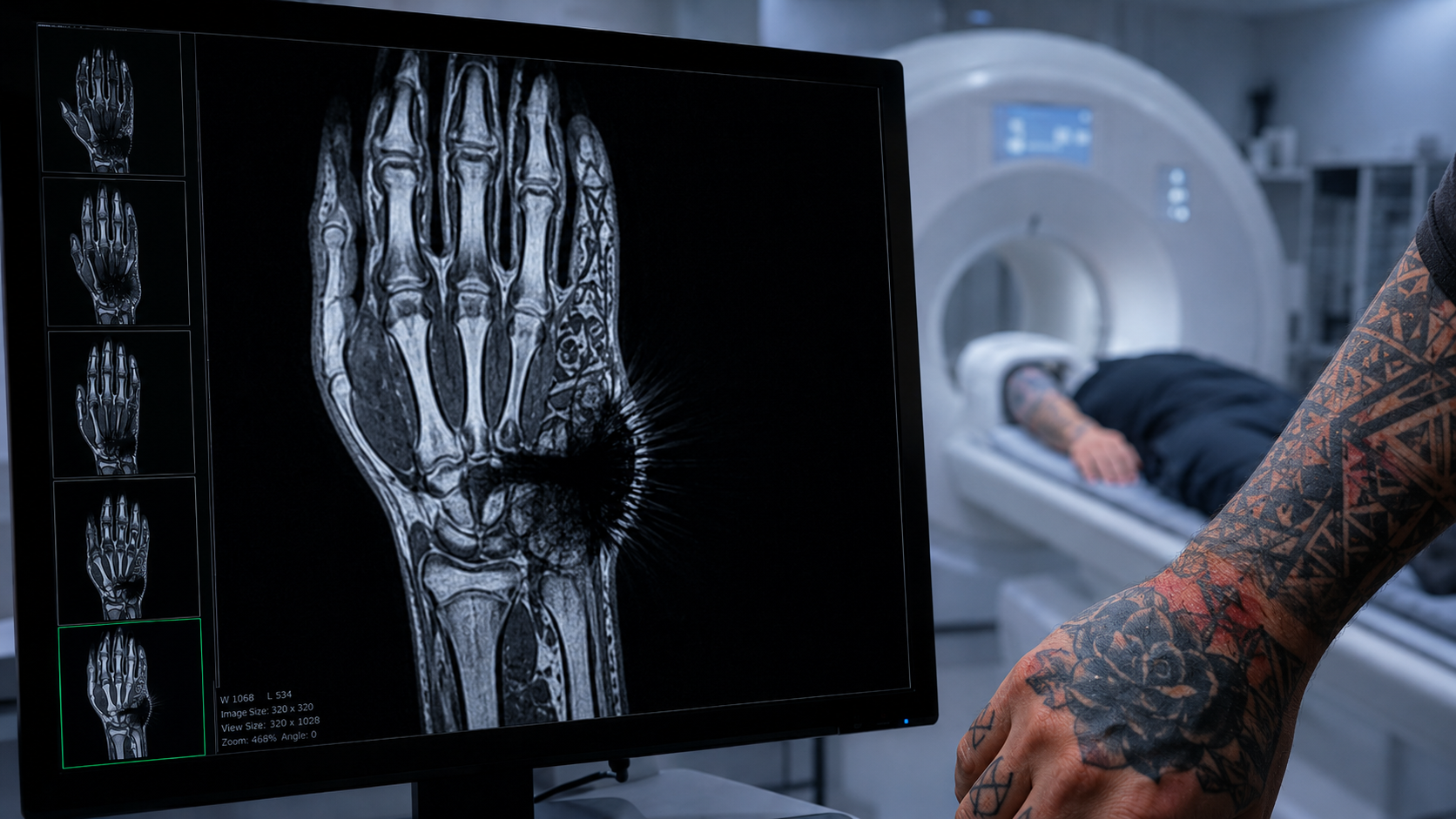
Carbon black and iron-oxide blacks create MRI signal voids. Mercury-sulfide reds and azo-dye compounds trigger persistent granulomatous reactions visible on ultrasound and CT scans.
USC Viterbi researchers harnessed tattoo pigment's unique optical fingerprints to barcode cancer-targeting nanoparticles — opening a new pathway for early tumour detection via Raman scanning.
The U.S. FDA gained authority over tattoo ink formulation only under MoCRA in late 2022. Decades of unregulated inks are now permanently embedded in the patient population passing through imaging departments every day.
Estimates from health surveys across developed nations suggest somewhere between 25 and 40 percent of adults carry at least one tattoo. That demographic is walking directly into imaging waiting rooms — and radiologists are increasingly navigating cases where ink is altering the diagnostic picture in ways that are not always immediately recognisable.
The problem is not that tattoos exist — it is that the medical imaging community has no standardised protocol for documenting or accounting for them.
On a plain chest radiograph, heavily tattooed areas over lung fields can introduce opaque patterns that mimic lesions. On MRI, ferromagnetic pigments create signal voids — dark patches where tissue data is simply absent. In breast imaging, nipple and chest-wall tattoos have been documented generating findings that triggered unnecessary patient callbacks, adding anxiety and cost to screening programmes.
Beyond image quality, there is also direct patient safety at stake during MRI. Certain metallic pigments — particularly iron oxides — can heat up or experience torque within a static magnetic field. While severe adverse events remain rare at standard clinical field strengths, the cumulative research points to a category of inks that warrant special attention in pre-scan screening protocols.
Nine out of ten commercially available U.S. inks carried significant discrepancies between label and actual chemical contents — a systemic gap that runs from the manufacturing floor to the scanning room, with no single point of accountability.
Without knowing what is in the bottle, neither the tattooist who applies the ink, the patient who wears it, nor the radiologist who scans through it can make fully informed decisions. The U.S. FDA gained authority to regulate tattoo inks only in late 2022 under the Modernization of Cosmetics Regulation Act — meaning decades of uncontrolled formulations are already permanently embedded in the patient population now passing through imaging departments every day.
The Chemistry Underneath the Skin
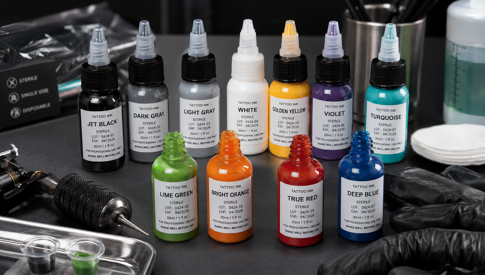
Tattoo pigments are not a single category of substance. They span inorganic metal salts, synthetic organic dyes, and carbon-based compounds — and each class behaves differently under imaging energy. Research from Amsterdam UMC, combining elemental and molecular analysis of both commercial inks and skin biopsies from patients with adverse reactions, found that pigments recovered from tissue are frequently chemically altered forms of what was originally injected. The body's immune response, UV exposure, and the mechanical trauma of deposition all modify ink chemistry over time.
Black inks — the most globally common pigment category — typically derive their colour from carbon black, the same fine particulate material used in printer cartridges and rubber tyres. Some formulations incorporate iron oxides, which are inherently magnetic. It is these iron-containing varieties that draw the most clinical concern in MRI settings. A 2024 murine study published in Experimental Dermatology confirmed that iron oxide pigment does not remain fixed at the injection site: it migrates via lymphatic channels to regional lymph nodes and ultimately concentrates in the liver. For clinical imaging, the footprint of a tattoo may extend far beyond the tattooed skin itself.
When the Scan Can't See Clearly
Research presented at the UK Imaging and Oncology Congress examined which specific ink compositions caused the greatest interference across imaging modalities. X-ray was found particularly vulnerable: high-density pigments attenuate radiation in ways similar to soft tissue abnormalities, creating shadow-like artefacts that trace skin contours. Experienced radiologists can often recognise these distribution patterns — but only when tattoo history has been communicated before the scan is acquired.
Red pigments occupy a separate category of clinical concern. Mercury sulfide (cinnabar), historically used in red and orange inks, is now regulated in parts of Europe but remains in circulation globally. Azo-based red dye substitutes carry their own profile of photodegradation products and have been implicated in persistent granulomatous reactions — nodular inflammatory responses that can appear as soft tissue abnormalities on ultrasound and CT.
Research from the University of Helsinki explored the biological persistence of tattoo pigment — how long these substances remain detectable in dermal tissue and how degradation products accumulate over years of residence. The findings reinforce the conclusion that the assumption of tattoo ink as a stable, cosmetically inert material is clinically outdated. Pigments break down, travel, and accumulate — all with consequences for how the human body appears under imaging scrutiny.
The Unexpected Opportunity
Not all of the science points toward risk. Researchers at USC Viterbi's Department of Biomedical Engineering took the same optical properties that make tattoo pigments detectable in tissue and turned them into a clinical asset. By attaching FDA-approved tattoo and food dye compounds to tumour-targeting nanoparticles, the team developed a new class of imaging contrast agents capable of illuminating cancer cells with greater specificity than conventional materials — with the critical advantage that the dyes carry existing regulatory approval.
The spectral signatures of tattoo pigments — their unique optical fingerprints detectable via Raman scanning — proved useful as identification barcodes for nanoparticles, allowing clinicians to differentiate malignant from healthy tissue with a precision not achievable through standard imaging alone. Published in Biomaterials Science, this work illustrates the dual nature of tattoo chemistry: simultaneously a source of diagnostic interference and a promising tool for imaging enhancement. The story of ink and medicine is not simply a cautionary tale — it is the story of a material injected into hundreds of millions of people without thorough scientific characterisation, which science is now racing to properly understand.
The Labelling Problem No One Fixed for Decades
Analysis of 54 commercially available inks from nine U.S. manufacturers, published in Analytical Chemistry, found that 45 — 83 percent — carried significant discrepancies between their stated ingredients and actual chemical contents. More than half contained undisclosed polyethylene glycol, a compound associated with organ damage under repeated exposure. Fifteen contained propylene glycol, a recognised allergen. One ink contained an antibiotic compound used to treat urinary tract infections; another contained 2-phenoxyethanol, which raises specific concerns for nursing infants.
Researchers could not determine whether unlisted ingredients were added intentionally or whether manufacturers had themselves been supplied with mislabelled raw materials — a supply chain opacity problem that amplifies risk at every level.
For imaging clinicians, this labelling gap is not merely a consumer safety issue — it is a live diagnostic complication. When an unexpected finding appears near a tattooed area, or when a patient develops a skin reaction following an MRI, clinicians need to know precisely what pigment compounds are present. If the product label cannot be trusted, the assumption that a given pigment is chemically inert cannot be trusted either. The FDA's authority under MoCRA arrived far too late to address the inks already embedded in patients entering imaging departments today.

Alsing KK et al. — Experimental Dermatology, 2024
Biodistribution of iron oxide tattoo pigment: An experimental murine study confirming lymph node and hepatic migration. PubMed PMID: 39304341. DOI: 10.1111/exd.15183
Amsterdam UMC — Tattoo Pigment Identification in Inks & Skin Biopsies
Complementary elemental and molecular bi-modal analysis of tattoo pigments in adverse reaction biopsies. Amsterdam UMC Pure portal. Peer-reviewed publication. pure.amsterdamumc.nl
Moseman K et al. — Analytical Chemistry, 2024 · Binghamton University / Swierk Lab
What's in my ink: An analysis of commercial tattoo ink composition on the U.S. market. DOI: 10.1021/acs.analchem.3c05687. 54 inks from 9 manufacturers examined.
Zavaleta C et al. — USC Viterbi / Biomaterials Science, 2020
Using FDA-approved coloring agents and tattoo dyes as optical contrast agents for surface-enhanced Raman spectroscopy nanoparticle-based cancer imaging. DOI: 10.1039/D0BM01099E
University of Helsinki — Tattoo Pigment Biological Persistence
Long-term dermal retention and degradation product accumulation of tattoo pigment compounds. Helsinki Digital Repository (Helda). Peer-reviewed. helda.helsinki.fi
UK Imaging and Oncology Congress — June 2026
Research presentation: differential imaging impact by tattoo ink type and composition across MRI, CT, X-ray, and ultrasound modalities. Congress proceedings, June 8, 2026.
This article draws on peer-reviewed studies, academic congress presentations, and university research communications. It is intended for general informational and editorial purposes and does not constitute medical advice. Patients with concerns about tattoo ink composition and upcoming medical imaging procedures should consult their referring physician or imaging provider before the scan.
All sources cited were independently reviewed and verified at the time of publication. Research findings referenced throughout represent the conclusions of the respective cited authors and institutions, and should be considered in the context of their full published works. Country-specific regulatory information may vary from general descriptions provided in this article.

Written by
MedBary Team
Comments
Log in to join the conversation.
Loading comments…